
Unraveling The Secrets Of Cell Division
By Emma Payne
During cell division, chromosomes are made up of two identical sister chromatids, which must be separated and pulled to opposite poles of the cell in order for the cell to divide properly. The structure responsible for moving the chromatids is the mitotic spindle, which is a complex structure composed of microtubules. These microtubules are anchored to the chromosomes at the kinetochore, which is a specialized protein complex located at the centromere of each chromosome.
The mitotic spindle is a dynamic structure that undergoes several changes during cell division. In early mitosis, the spindle forms between the two poles of the cell and the microtubules begin to attach to the kinetochores of the chromosomes. As mitosis progresses, the spindle fibers shorten, pulling the chromosomes to the poles of the cell. Once the chromosomes reach the poles, the spindle fibers disassemble and the chromosomes decondense, returning to their normal interphase state.
The mitotic spindle is essential for the proper segregation of chromosomes during cell division. If the spindle is not properly formed or if the microtubules are not properly attached to the kinetochores, the chromosomes will not be able to separate correctly and the cell will not be able to divide.
What Moves the Chromatids During Cell Division
During cell division, chromosomes are made up of two identical sister chromatids, which must be separated and pulled to opposite poles of the cell in order for the cell to divide properly. The structure responsible for moving the chromatids is the mitotic spindle, which is a complex structure composed of microtubules. These microtubules are anchored to the chromosomes at the kinetochore, which is a specialized protein complex located at the centromere of each chromosome.
- Mitosis: The process of cell division that results in two daughter cells with the same number of chromosomes as the parent cell.
- Chromatid: One of the two identical copies of a chromosome that are formed during DNA replication.
- Kinetochore: A specialized protein complex located at the centromere of each chromosome that serves as the attachment point for microtubules during cell division.
- Microtubule: A long, thin, hollow cylinder composed of tubulin protein subunits that forms part of the cytoskeleton and is involved in various cellular processes, including cell division.
- Mitotic spindle: A complex structure composed of microtubules that forms during cell division and is responsible for separating and pulling the chromatids to opposite poles of the cell.
- Centromere: The region of a chromosome where the two sister chromatids are joined together.
- Kinetochore fiber: A microtubule that attaches to the kinetochore of a chromosome during cell division.
- Polar fiber: A microtubule that extends from one pole of the cell to the other and does not attach to a kinetochore.
The mitotic spindle is essential for the proper segregation of chromosomes during cell division. If the spindle is not properly formed or if the microtubules are not properly attached to the kinetochores, the chromosomes will not be able to separate correctly and the cell will not be able to divide.
Mitosis
Mitosis is a critical process in the life of a cell, as it allows for the creation of two new cells with the same genetic material as the parent cell. This process is essential for growth, development, and reproduction. Mitosis is also essential for the repair of damaged tissues.
The movement of chromatids during cell division is a key part of mitosis. Chromatids are the individual strands of DNA that make up chromosomes. During mitosis, the chromatids must be separated and pulled to opposite poles of the cell in order for the cell to divide properly. The structure responsible for moving the chromatids is the mitotic spindle, which is a complex structure composed of microtubules. These microtubules are anchored to the chromosomes at the kinetochore, which is a specialized protein complex located at the centromere of each chromosome.
The mitotic spindle is essential for the proper segregation of chromosomes during cell division. If the spindle is not properly formed or if the microtubules are not properly attached to the kinetochores, the chromosomes will not be able to separate correctly and the cell will not be able to divide. This can lead to a number of problems, including cell death, birth defects, and cancer.
The study of mitosis and the movement of chromatids during cell division is an important area of research. This research has led to a better understanding of the cell cycle and the development of new treatments for cancer and other diseases.
Chromatid
Chromatids are the individual strands of DNA that make up chromosomes. During cell division, the chromatids must be separated and pulled to opposite poles of the cell in order for the cell to divide properly. The structure responsible for moving the chromatids is the mitotic spindle, which is a complex structure composed of microtubules. These microtubules are anchored to the chromosomes at the kinetochore, which is a specialized protein complex located at the centromere of each chromosome.
- The structure of chromatids
Chromatids are composed of DNA and proteins. The DNA is organized into a series of nucleosomes, which are protein-DNA complexes. The nucleosomes are arranged in a repeating pattern along the length of the chromatid.
- The role of chromatids in cell division
Chromatids are essential for cell division. During mitosis, the chromatids are separated and pulled to opposite poles of the cell. This ensures that each daughter cell receives a complete set of chromosomes.
- The importance of chromatids
Chromatids are essential for the proper development and function of organisms. If the chromatids are not properly separated during cell division, it can lead to a number of problems, including birth defects, cancer, and cell death.
The study of chromatids and their role in cell division is an important area of research. This research has led to a better understanding of the cell cycle and the development of new treatments for cancer and other diseases.
Kinetochore
The kinetochore is a specialized protein complex located at the centromere of each chromosome that serves as the attachment point for microtubules during cell division. It plays a critical role in ensuring the proper segregation of chromosomes during cell division.
- Structure of the kinetochore
The kinetochore is a large protein complex that is composed of more than 100 different proteins. It is organized into a series of subcomplexes, each of which has a specific function.
- Function of the kinetochore
The kinetochore serves as the attachment point for microtubules during cell division. Microtubules are long, thin protein filaments that form part of the mitotic spindle, which is the structure that pulls the chromosomes to opposite poles of the cell.
- Regulation of the kinetochore
The kinetochore is regulated by a complex network of proteins. These proteins ensure that the kinetochore is properly attached to the microtubules and that the chromosomes are properly segregated during cell division.
- Kinetochore and cancer
Defects in the kinetochore can lead to cancer. These defects can result in the missegregation of chromosomes, which can lead to the development of cancer cells.
The kinetochore is a critical structure for cell division. It ensures that the chromosomes are properly segregated during cell division, which is essential for the proper development and function of organisms.
Microtubule
Microtubules are long, thin protein filaments that form part of the cytoskeleton of cells. They are involved in a variety of cellular processes, including cell division. During cell division, microtubules form the mitotic spindle, which is the structure that pulls the chromosomes to opposite poles of the cell.
- Structure of microtubules
Microtubules are composed of a protein called tubulin. Tubulin molecules are arranged in a repeating pattern to form a hollow cylinder. Microtubules are highly dynamic structures that can quickly assemble and disassemble.
- Function of microtubules in cell division
Microtubules play a critical role in cell division. They form the mitotic spindle, which is the structure that pulls the chromosomes to opposite poles of the cell. The mitotic spindle is composed of two sets of microtubules: kinetochore microtubules and polar microtubules. Kinetochore microtubules attach to the chromosomes at the kinetochore, which is a specialized protein complex located at the centromere of each chromosome. Polar microtubules extend from one pole of the cell to the other and overlap with the kinetochore microtubules.
- Regulation of microtubules
The assembly and disassembly of microtubules is regulated by a complex network of proteins. These proteins ensure that the microtubules are properly formed and that they are attached to the chromosomes at the correct time.
- Microtubules and cancer
Defects in microtubules can lead to cancer. These defects can result in the missegregation of chromosomes, which can lead to the development of cancer cells.
Microtubules are essential for cell division. They play a critical role in ensuring that the chromosomes are properly segregated during cell division, which is essential for the proper development and function of organisms.
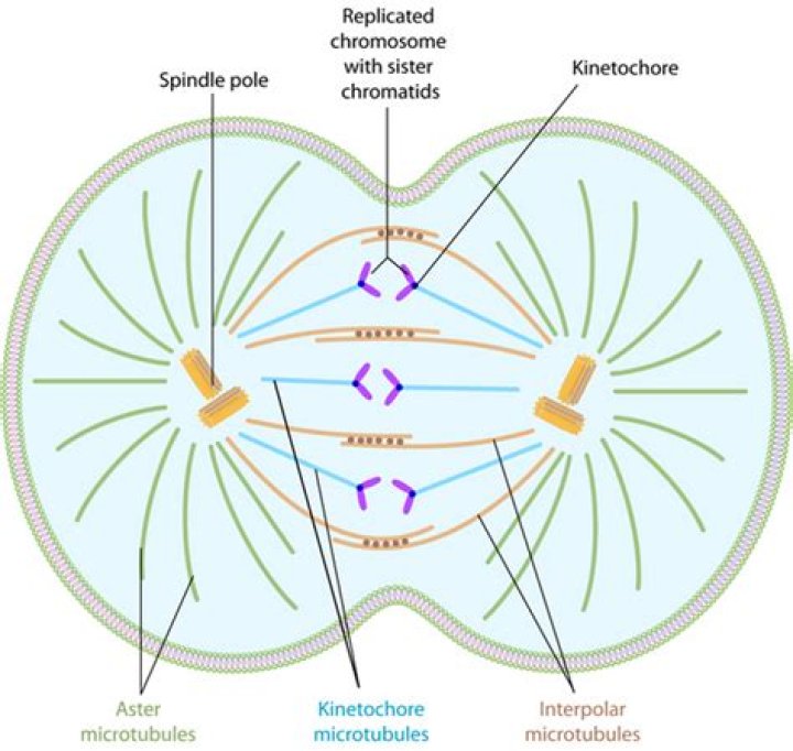
Mitotic spindle
The mitotic spindle is a complex structure composed of microtubules that forms during cell division and is responsible for separating and pulling the chromatids to opposite poles of the cell. It is a key component of the "what moves the chromatids during cell division what organelle anchors these" complex, which also includes the kinetochore and the centromere.
The mitotic spindle forms during prophase, the first stage of mitosis. It is composed of two sets of microtubules: kinetochore microtubules and polar microtubules. Kinetochore microtubules attach to the chromosomes at the kinetochore, a specialized protein complex located at the centromere of each chromosome. Polar microtubules extend from one pole of the cell to the other and overlap with the kinetochore microtubules.
Once the mitotic spindle is formed, it begins to shorten. This shortening pulls the chromosomes to the poles of the cell. As the chromosomes reach the poles, the mitotic spindle disassembles and the chromosomes decondense, returning to their normal interphase state.
The mitotic spindle is essential for the proper segregation of chromosomes during cell division. If the mitotic spindle is not properly formed or if the microtubules are not properly attached to the kinetochores, the chromosomes will not be able to separate correctly and the cell will not be able to divide. This can lead to a number of problems, including birth defects, cancer, and cell death.
The study of the mitotic spindle and its role in cell division is an important area of research. This research has led to a better understanding of the cell cycle and the development of new treatments for cancer and other diseases.
Centromere
The centromere is a specialized region of a chromosome that is essential for cell division. It is the region where the two sister chromatids, which are identical copies of each other, are joined together. The centromere also serves as the attachment point for the mitotic spindle, which is the structure that pulls the chromosomes to opposite poles of the cell during cell division.
The centromere is a complex structure that is composed of DNA and proteins. The DNA in the centromere is organized into a series of repetitive sequences, which are essential for the binding of the proteins that make up the mitotic spindle. The centromere is also the site of attachment for the kinetochore, which is a large protein complex that serves as the attachment point for the microtubules of the mitotic spindle.
The centromere is essential for the proper segregation of chromosomes during cell division. If the centromere is not properly formed or if the mitotic spindle is not properly attached to the centromere, the chromosomes will not be able to separate correctly and the cell will not be able to divide. This can lead to a number of problems, including birth defects, cancer, and cell death.
The study of the centromere and its role in cell division is an important area of research. This research has led to a better understanding of the cell cycle and the development of new treatments for cancer and other diseases.
Kinetochore fiber
Kinetochore fibers are essential for the proper segregation of chromosomes during cell division. They attach to the kinetochores of chromosomes and pull them to opposite poles of the cell. This ensures that each daughter cell receives a complete set of chromosomes.
Kinetochore fibers are composed of microtubules, which are long, thin protein filaments. Microtubules are highly dynamic structures that can quickly assemble and disassemble. This allows the kinetochore fibers to lengthen and shorten as needed to ensure that the chromosomes are properly segregated.
The attachment of kinetochore fibers to the kinetochores is mediated by a complex of proteins known as the kinetochore. The kinetochore is a large, multi-protein structure that serves as the attachment point for the mitotic spindle. The mitotic spindle is the structure that pulls the chromosomes to opposite poles of the cell.
The proper attachment of kinetochore fibers to the kinetochores is essential for the proper segregation of chromosomes during cell division. If the kinetochore fibers are not properly attached, the chromosomes will not be able to separate correctly and the cell will not be able to divide. This can lead to a number of problems, including birth defects, cancer, and cell death.
The study of kinetochore fibers and their role in cell division is an important area of research. This research has led to a better understanding of the cell cycle and the development of new treatments for cancer and other diseases.
Polar fiber
Polar fibers are a type of microtubule that extends from one pole of the cell to the other and does not attach to a kinetochore. They are part of the mitotic spindle, which is the structure that pulls the chromosomes to opposite poles of the cell during cell division.
Polar fibers play an important role in cell division by helping to align the chromosomes and ensuring that they are properly segregated into the two daughter cells. They also help to maintain the shape of the mitotic spindle and to prevent the chromosomes from becoming tangled.
The proper function of polar fibers is essential for the accurate segregation of chromosomes during cell division. Defects in polar fibers can lead to a number of problems, including aneuploidy, which is a condition in which cells have an abnormal number of chromosomes. Aneuploidy can lead to a variety of developmental problems and diseases, including cancer.
The study of polar fibers and their role in cell division is an important area of research. This research has led to a better understanding of the cell cycle and the development of new treatments for cancer and other diseases.
FAQs about "what moves the chromatids during cell division what organelle anchors these"
This FAQ section provides concise answers to common questions about "what moves the chromatids during cell division what organelle anchors these".
Question 1: What are chromatids?
Chromatids are the individual strands of DNA that make up chromosomes. During cell division, the chromatids must be separated and pulled to opposite poles of the cell in order for the cell to divide properly.
Question 2: What is the mitotic spindle?
The mitotic spindle is a complex structure composed of microtubules that forms during cell division and is responsible for separating and pulling the chromatids to opposite poles of the cell.
Question 3: What is the kinetochore?
The kinetochore is a specialized protein complex located at the centromere of each chromosome that serves as the attachment point for microtubules during cell division.
Question 4: What is the centromere?
The centromere is the region of a chromosome where the two sister chromatids are joined together.
Question 5: What is the role of microtubules in cell division?
Microtubules are long, thin protein filaments that form part of the cytoskeleton of cells. They are involved in a variety of cellular processes, including cell division. During cell division, microtubules form the mitotic spindle, which is the structure that pulls the chromosomes to opposite poles of the cell.
Question 6: What happens if the mitotic spindle is not properly formed or attached to the chromosomes?
If the mitotic spindle is not properly formed or attached to the chromosomes, the chromosomes will not be able to separate correctly and the cell will not be able to divide. This can lead to a number of problems, including birth defects, cancer, and cell death.
Summary
The movement of chromatids during cell division is a complex process that is essential for the proper development and function of organisms. The mitotic spindle, kinetochore, centromere, and microtubules all play important roles in this process. Defects in any of these components can lead to a number of problems, including birth defects, cancer, and cell death.
Transition
The study of cell division is an important area of research. This research has led to a better understanding of the cell cycle and the development of new treatments for cancer and other diseases.
Tips for understanding "what moves the chromatids during cell division what organelle anchors these"
Understanding the movement of chromatids during cell division is essential for students of biology and medicine. Here are a few tips to help you master this complex topic:
Tip 1: Start with the basics. Before you can understand the movement of chromatids, you need to have a solid foundation in the basics of cell division. This includes understanding the different stages of cell division, the structure of chromosomes, and the role of the mitotic spindle.
Tip 2: Focus on the key players. The movement of chromatids is driven by a complex interplay of proteins and organelles. The most important players to focus on are the mitotic spindle, the kinetochore, and the centromere.
Tip 3: Use visual aids. Diagrams and animations can be very helpful for visualizing the movement of chromatids. There are many resources available online and in textbooks that can provide you with these visual aids.
Tip 4: Practice, practice, practice. The best way to master this topic is to practice solving problems. There are many practice problems available online and in textbooks.
Tip 5: Ask for help. If you are struggling to understand this topic, don't be afraid to ask for help. Your teacher, professor, or a tutor can help you to clarify the concepts and answer your questions.
Summary
By following these tips, you can improve your understanding of the movement of chromatids during cell division. This is an essential topic for students of biology and medicine.
Transition
The movement of chromatids during cell division is a complex and fascinating process. By understanding this process, you can gain a deeper appreciation for the beauty and complexity of life.
Conclusion
The movement of chromatids during cell division is a complex and essential process that ensures the accurate segregation of chromosomes. This process is driven by a complex interplay of proteins and organelles, including the mitotic spindle, the kinetochore, and the centromere. Defects in any of these components can lead to a number of problems, including birth defects, cancer, and cell death.
The study of cell division is an important area of research that has led to a better understanding of the cell cycle and the development of new treatments for cancer and other diseases. By continuing to study this process, we can gain a deeper understanding of the fundamental mechanisms of life.
Uncover The Secrets Behind Phil Murphy's Teeth: A Dental Deep Dive
Uncover Jonathan Banks' Residence: Exploring His Roots And Lifestyle
Betty Gabriel: A Rising Star Unlocking Boundaries And Inspiring Dreams